3 Ways the Incentives are Misaligned for Training Pathology Residents

carrotI enjoy teaching residents and believe that helping those who are struggling is possibly the greatest impact we can have. Whereas in our clinical work we help one patient at a time, teaching residents allow us to help exponentially more patients by extending our expertise geographically outward and temporally forward. I have noticed, however, that the effort required to train residents is not incentivized. Here are 3 reasons why many attendings at academic institutions have little interest in expending anything beyond the minimum effort required to train residents:

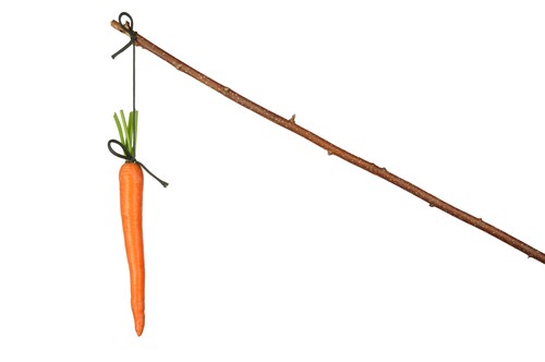
  1. It is very unusual these days for a graduating AP/CP resident to find a job in the absence of one or two subsequent year-long subspecialty fellowships. Awareness of this fact means that attendings do not feel as strong a sense of urgency to ensure that their residents hone their clinical skills sufficiently before they graduate, since they will have more time after they graduate before they apply for a job. At that point it will be someone else’s responsibility.
  2. Residents are given the opportunity to evaluate their attendings in an anonymous fashion. Negative evaluations affect our ability to be promoted academically. This matters because academic level affects our reputation and salary. Many attendings have found that the easiest way to get good evaluations is to keep their mouths shut when residents are under-performing and to take the work themselves. Trying to help a resident who is under-performing can be emotionally taxing and even risky. First, you will inevitably hurt their feelings, no matter how gently you broach the subject and however well you “sandwich” the negative aspects between protective layers of positive feedback. Many of us don’t like hurting people’s feelings in the short-term, even if it will help them succeed in the long term. Second, while some residents appreciate the effort and risk that attendings take in helping them remediate their skills – others retaliate – and what better forum for retaliation than an anonymous online system.
  3. Academic promotion (at least in my institution and probably many others) also depends on teaching medical students. We are expected to log every teaching activity with the medical students, and each type of teaching activity is assigned a certain number of points.  On the other hand, while we are tacitly expected to teach residents while on service, there are no repercussions for abrogating this responsibility. If we tell the residents that they are doing a good job and give them good evaluations (i.e. quickly click on high scores in an online form), then often no one will complain. Medical student education is therefore prized more than residency education, despite the fact that teaching pathology to medical students probably has little importance beyond helping them pass Step 1 of their boards.

Of course, it is easier to identify problems than it is to propose solutions. In the future, I hope to write about ways we can incentivize quality pathology residency teaching.

I’d love to hear your thoughts.


Too many pathology societies for our own good?

With more challenges to our profession from new technologies (machine vision, circulating DNA, in vivo microscopy) along with decreased reimbursement, we need to ensure that we stay relevant to clinical medicine.

dilutionOur various societies are led by smart and driven people who understand this need, now more than ever. However it seems to me that we have too many societies which duplicate each other’s efforts and weaken our overall capacity to advocate.

In particular, I would propose merging the 3 big societies of the United States and Canadian Academy of Pathology (USCAP), the College of American Pathologists (CAP), and the American Society of Clinical Pathology (ASCP). I understand that the USCAP has more of an academic bent, the CAP’s emphasis is on professional advocacy for pathologists, and the ASCP caters more to laboratory medicine. However, I believe that these different functions can be combined together into one professional organization for pathology professionals in the United States.

Supporting this proposal is the existence of overlap in the functions of these different organizations, especially in the realms of research and education. At the same time since the different groups cater to different elements of our profession (e.g. USCAP to those in academic practice, CAP to community/private practice, and ASCP to laboratory medicine professionals and technologists), it weakens our ability to make decisions as a cohesive unit.

In a previous era, a plethora of various societies with different leadership and political aims worked fine. But in today’s cost-conscious environment, every time I am separately asked for membership fees to the three above organizations, I cannot help but wonder if we are diluting ourselves to the point of irrelevance.

[Photo courtesy of http://www.wikiwand.com/fr/Dilution]

Pathology is now cool at the lectern

I had the sorry experience recently of trying to teach some basic upper GI tract pathology to a group of 4th year medical students going into surgery, by using a PowerPoint presentation, only to watch many of them glaze over and start peeking every few moments at their phones.

james deanIt may be that I don’t give good PowerPoint presentations, but I think there is something about PowerPoint that makes it difficult to convey the dynamic nature of tissue interpretation. This is problematic because many medical students do not rotate through Anatomic Pathology and thus don’t really have any idea what we actually do. Worse, even, they may assume that our work is boring – meaning that we will miss out on recruiting smart medical students for whom our specialty would be a great fit.

Shortly after this experience I was thinking about what I could do differently next time and learned about PathPresenter. Apparently the creators of this platform had also been contemplating a better way to convey histopathology to an audience, and they created a masterful solution.

PathPresenter allows you to basically embed whole slide images (WSIs) into a presentation and then select fields of interest from the WSI to show. However, what I love most is that within the presentation, you can move the whole slide image around and zoom in and out. This way you can weave educational slides with dynamic WSI’s seamlessly. You can upload your own WSI files if you want, or you can use the extensive library of WSI’s that the creators of PathPresenter have built and are constantly enlarging.

The platform is free to use and I am excited to start using it, especially the next time I am asked to give a lecture to medical students or to explain to a non-pathologist “what is it again that you do?”

My answer next time will be, “Watch, I’ll show you.”


What happens when pathologists talk with clinicians…

I enjoyed this anecdote from my colleague Dr. Keith Kaplan. It reminded me of Joseph Heller’s Catch 22.

A Conversation with a Clinician

Here’s a clip:

Dr. [Clinician]: When I biopsied the patient before, the mucosa looked funny.  You said it looked atrophic.

Me: Yes.

Dr. [Clinician]: The mucosa looked funny.

Me: How did the mucosa look funny?

Dr. [Clinician]: You know, funny.

Me: No I don’t know. You said the mucosa looked funny. We don’t have a diagnosis for “funny”.

Dr. [Clinician]: You think this is funny? How is this funny?

Me: No. This is not funny. You said the mucosa looked funny. I was worried about a carcinoma.

Dr. [Clinician]: Yeah, the mucosa looked funny.

Read more: http://tissuepathology.com/2017/03/14/an-conversation-with-a-clinician/#ixzz4bRXVp8vS
Under Creative Commons License: Attribution
Follow us: @tissuepathology on Twitter | pathology2.0 on Facebook

M8: A microscope on steroids

At the USCAP 2017 conference in San Antonio, I had a chance to try out the Precipoint M8 (http://www.precipoint.com/m8-microscope), which is a combined microscope and slide scanner. It is an entirely new concept and is the only example I am aware of on the market. It starts by scanning a low-power overview of the slide after which the user can zoom in and examine the slide at higher power. As the user zooms in or pans around, the M8 quickly scans multiple fields of view and stitches them together on the screen. Since the slide remains on the stage, the user can adjust the focus (unlike other whole slide imagers which scan at a single focal plane typically). Using a large high-resolution monitor, one essentially has a much larger field of view than one would have with a regular microscope without loss of z-axis information. The user can also scan the entire slide or a specific area of the slide to be stored as a file.

A critic’s first response might be, “Why would you want a real time slide scanner when you can just scan the slides in advance with a whole slide imager?” I would suggest 3 responses:

1. A real time slide scanner such as the M8 allows the user to adjust the focus continuously. Although other slide scanners are starting to provide the option of scanning in 2 or more predetermined focal planes, discrete planes of focus inherently provide less information than exists on an actual slide with several microns worth of continuous z-axis information.

2. Scanning the slides in advance means the physical slides are delivered to the pathologist later. When my department at UPMC was trying out the Omnyx system during pilot testing, most of us felt that we needed to still examine the original slides to make sure we weren’t missing information that was lost by scanning only a single focal plane. Of course we examined the original slides anyway since digital pathology was not and still is not FDA approved for primary diagnosis, but most of us felt that we still would have liked to examine the original slides even if we were not required to. Therefore, we felt like there was little to gain by trying to interpret whole slide images and were discouraged that we received our slide later.

3. Accessing previously scanned whole slide images requires downloading them from a server, which can incur a delay depending on the available bandwidth at one’s institution.

Now the M8 isn’t better than a microscope in every way. With a microscope, the user experiences no delay when changing the magnification or panning the image, even though the field of view is smaller. It is interesting to wonder how much faster this technology can theoretically be pushed – if the delay in filling in those squares on the screen can be reduced to the point where it is no longer perceptible. Then I would consider ditching my microscope (if the price is right).

Does Pathologists’ Physical Presence in the Hospital Matter?

houses
From speaking with my colleagues, it seems like there is a trend of pathology departments at academic medical centers moving out of the main hospital and into separate buildings. Sometimes their new quarters are a significant distance from the hospital, including the operating rooms. Hospital administrators prefer to use the space for more direct patient services such as additional inpatient beds, and assume that pathologists can do their jobs remotely with the aid of digital imaging, much like radiologists. Some pathologists, especially those who are enthusiastic about digital pathology, embrace this challenge as an opportunity to adopt new technologies. Others are skeptical about whether they can do their jobs as well and as quickly without the physical slides in their hands.

However, the biggest push back seems to be from surgeons. Many of them are uncomfortable knowing that there is no pathologist present on site with whom they can discuss and orient gross specimens or intraoperative findings in person. Different surgeons show varying levels of interest in our work, but there is certainly a significant proportion who seem to worry that the lack of in-person interaction will erode our professional relationship and lead to inferior patient care.

In my experience, surgeons are especially sensitive to the level of confidence that they perceive in pathologists (and in each other). If they sense a lack of confidence, as is often the case with junior pathologists straight out of training, they may ask for second opinions of more senior pathologists whom they know better. I think that part of their discomfort with our being off site is that our interactions with them are as much about the nuances of how we project confidence as about the content of our diagnoses, especially for frozen sections. It will be more difficult for them to “read” us if they don’t know how we interact with people and how we sound when we are confident.

In our eagerness to apply new technologies to our practice and push our field into the future, it is somewhat jarring to realize that many of our colleagues really just want to have us around. Perhaps we should feel honored!

[The picture was borrowed from Vista Window Film Blog.]

How should we teach pathology to medical students?

apple
Pathology, translated etymologically means “the study of disease.” The word carries a lot of weight. After all, “the study of disease” is the foundation and inspiration of basically all of medicine and biomedical science. The desire and effort to understand how and why diseases occur has probably existed for as long as civilization.

Although the pure etymological definition of pathology is the study of disease, most pathologists don’t actually spend their time studying diseases. We study tissue, to  diagnose diseases and identify prognostic features. The fact that we are still called pathologists dates to when microscopic examination of affected tissue was essentially the only way of studying and characterizing diseases beyond history and physical examination.

Many diseases are still diagnosed by their microscopic features, cancer being perhaps the most prominent example. But since the days of Rudolph Virchow, the mechanisms of many diseases which were previously characterized only microscopically became better understood. With the acceptance of the germ theory of disease and with developments in biochemistry and immunology, pathophysiology could be explained by mechanisms which are often not apparent at the microscopic level.

The historical preeminence of histology in the characterization of diseases lives on today in the way medical students are educated. The fact that pathology is a core discipline of medical school pre-clinical education (years 1 and 2) reflects an acceptance among physician educators that the appearance of diseased tissue at the microscopic level, in the realm of about 40x-400x, after being fixed in formalin, embedded in paraffin, sectioned at approximately 7 micron thickness, and colorized with pink and blue stains, contains a fundamental truth about what a disease essentially is.

Of course, only those medical students who will specialize in pathology will actually interpret histopathology once they are physicians. Perhaps this is why pathology has become less emphasized in many medical schools compared to its prominence in the past. Nonetheless, it remains entrenched in the curriculum, and Robbins Basic Pathology remains an essential text for medical students. Despite this emphasis during medical school on teaching classic histologic appearances of diseases, most students graduate medical school without having much of a clue about what pathologists actually do, what real microscopic interpretation entails, and what information pathologists can provide.

Perhaps pathologists involved in medical school education and USMLE Step 1 test development should focus more on understanding principles of specimen evaluation and terminology of pathology reports, to facilitate professional interaction and communication in the future. While only those who pursue a pathology residency will interpret tissue at the microscopic level, almost all physicians will rely on pathology reports and conversations with pathologists to help manage their patients.

[Photo is courtesy of the Huffington Post: http://www.huffingtonpost.ca/kathy-buckworth/teacher-gifts_b_5521055.html]

2 possible futures for pathology

Here is one possible future: Some day, whole slide imagers will be ubiquitous in pathology labs throughout the U.S. They will scan our slides quickly and in multiple contiguous focal planes, thus retaining all the morphologic data that exists in “analog” form. The quality of the whole slide imaging will be so good that pathologists will have no need to review the original slides. Our workflow will be entirely digital and physical proximity of the histology lab and the pathologist will no longer matter. Artificial intelligence algorithms will interpret the scanned slides and will flag the urgent diagnoses. At first computer assisted diagnosis will make pathologists’ jobs easier and we will be able to do more work faster and extract more information from morphology than we could before. However, eventually pathologists will have less to do as the computers become smarter and faster. At some point slide interpretation will no longer be done by human beings, since AI can do everything they do faster and are less prone to error. Furthermore, they will learn to identify complexnexi patterns with diagnostic and prognostic relevance which are beyond human perception. Patterns such as chromatin distribution, reconstructed 3D tissue architecture, and quantification of nuclear contour irregularity.

Here is another possible future: In vivo microscopy will improve to the point where it matches the resolution of histology, thus precluding the need for ex vivo microscopic interpretation. In conjunction with improvements in metabolic imaging and exosomal DNA analysis, histology will have nothing to contribute beyond the information which can be obtained in vivo. Whole slide imaging will become a redundant concept since there will no longer be slides to scan. Pathologists, radiologists, and clinicians who do endoscopy will compete for the professional right to interpret these in vivo images which will render most biopsies unnecessary.

Will one of these scenarios prevail? Or will the future be different?

[Photo is of Nexi, a robot developed at MIT]

Part 2: Will Pathology Merge with Radiology?

puzzlepieceI was taken by surprise when I read the recent editorial in JAMA which casually suggested a merging of the disciplines of pathology and radiology, and I voiced my skepticism. Since then I have read more on the subject and learned that this is not a new idea. The proposed discipline which would encompass radiology, pathology, and molecular testing has variously been called “diagnostic medicine,” “integrated diagnostics,” and “full service diagnostics,” among other terms.

The CEO of General Electric, Jeff Immelt, and Dave Hickey of Siemens have expressed their support of this concept, the former in 2012 and the latter in 2007.

Interestingly the most convincing case for the merging of pathology and radiology was actually made by a prominent pathologist, Dr. Bruce Friedman, in 2006, on his blog Lab Soft News. In that post he presented “Ten Reasons for merging pathology/lab medicine with radiology.” The full text can be read by following the above link but in my opinion the strongest reasons (partly edited for brevity) are these five:

  1. Both share a mission of diagnosing disease through images and biomarkers.
  2. The integrated reports of pathologists and radiologists working collaboratively would achieve higher levels of quality.
  3. The science and research agendas of molecular imaging and molecular diagnostics already demonstrate extensive overlap.
  4. Much of the core technologies used in surgical pathology are badly outdated and would benefit from an infusion of the new science and technology currently being explored in medical imaging and molecular imaging.
  5. Pathology and lab medicine need a greater influx of capital investment in the form of corporate R&D funds analogous to that supporting molecular imaging.

My assessment is that there are different levels to what is meant by a “merging” of these disciplines. I will order these by increasing levels of difficulty to implement. The first level would be integration of our reports into a single pathology-radiology report, which can be achieved fairly easily with software and which is already done at UCLA. The next level is integration of our physical facilities and administrative help, for which the barriers would be mostly political and administrative. The third level, which is admittedly a huge jump from the last step, would be the merging of our specialties into an integrated knowledge base, a single professional identity, and a revamped residency training.

While the latter is practically inconceivable at this point given the profound differences in our respective knowledge bases, the first two levels of integration are entirely within the realm of plausibility. In fact many pathologists and radiologists may welcome the possibility of working more closely as an opportunity to enhance their clinical expertise.

As for the third level of integration, an actual merging of our disciplines, radiologists are probably farther along in considering this than pathologists. While artificial intelligence (AI) poses an imminent threat to radiologists, pathologists are less concerned for now, because we have witnessed efforts to adopt digital pathology fail. And without routine digitization of our microscopic slides, AI will not get a foothold. It is still difficult at this point to make a strong business case for adopting digital pathology. To my perspective, that radiologists are talking more frequently and more publicly about merging with pathology is an effort to stay relevant in the face of a serious threat to their specialty. Pathologists will eventually feel a similar threat once the financial and technical barriers to digitization decrease and AI advances to the point where it can perform complex image interpretation. In the meantime, though, it would behoove both of our professions to begin this dialogue together

Will Pathology Merge with Radiology?

merge-signIn an editorial just published in JAMA, Drs. Eric Topol (director of the Scripps Translational Science Institute) and Saurabh Jha (a radiologist at University of Pennsylvania) propose the merging of pathology and radiology into a single “information specialist” discipline. They argue that since computers will be able to perform the manual aspects of image interpretation, humans can focus on higher tasks without the need for training in these nuances.

The details of their proposed merger are not fleshed out fully but the general concept is that these information specialists would:

interpret the important data, advise on the added value of another diagnostic test, such as the need for additional imaging, anatomical pathology, or a laboratory test, and integrate information to guide clinicians.

Their proposal is non-specific enough that it is difficult to critically assess. Notice how blithely they propose that these information specialists will “interpret the important data” without further elaboration. What constitutes “important”? New diagnoses of malignancy? Identification of infectious organisms? Positive margins?

A problem with this suggestion, however non-specific, is that one wonders how these specialists will be able to “interpret the important data,” whatever it is exactly, when their training in morphologic interpretation is so diluted:

Information specialists should train in the traditional sciences of pathology and radiology. The training should take no longer than it presently takes because the trainee will not spend time mastering the pattern recognition required to become a competent radiologist or pathologist. Visual interpretation will be restricted to perceptual tasks that artificial intelligence cannot perform as well as humans. The trainee need only master enough medical physics to improve suboptimal quality of medical images. Information specialists should be taught Bayesian logic, statistics, and data science and be aware of other sources of information such as genomics and biometrics, insofar as they can integrate data from disparate sources with a patient’s clinical condition.

Thus, within the same length of time as it currently takes to become adept in either pathology or radiology, these new specialists will train in both fields and also become adept in Bayesian logic, statistics, and data science. Without being well-versed in the minutiae of image interpretation, will these future specialists be able to perform higher level perceptual tasks that AI cannot perform as well? I’m not sure I would trust them.

However, there is merit to some of their argument. Machine learning can in some instances predict patients’ prognoses better than pathologists can. As shown in a recent study they cite, machine learning can predict prognosis of non-small cell lung carcinoma patients more accurately than our current grading and staging criteria, using image analysis parameters that can only be discovered and quantified by software. Thus in the future we may scrap our current grading and staging schemes as machine learning combined with molecular testing better predicts patient prognoses and responses to therapy.

At some point in the future, AI will no doubt displace the skills of the pathologist, but by then it will be supplanting many other medical specialties which similarly will be more competently performed by AI. In the near term, though, replacing specialized training in pathology and radiology would leave no one – neither humans nor computers – who is competent to interpret the important data.

Finally, aside from the text of this piece, what is perhaps even more illuminating is that the authors of a proposal in a leading medical journal to completely revamp the practice of pathology and merge it with another did not even include a pathologist. We as a specialty are doing something wrong if this conversation is taking place without us.